Medial Malleolus Ankle Fracture The Radiology Assistant Ankle

The Radiology Assistant Special Ankle Fractures Publicationdate 2012 08 23. classification of ankle fractures is important in order to estimate the extent of the injury and the stability of the joint. the weber classification focuses on the integrity of the fibula and the syndesmosis, which holds the ankle mortise together. the lauge hansen system focuses on the trauma mechanism. There are two positions of the foot in which the flexible ankle joint becomes a rigid and vulnerable system: extreme supination and pronation. in these positions forces applied to the talus within the ankle mortise can result in fractures of the malleoli and rupture of the ligaments. in 80% of ankle fractures the foot is in supination.

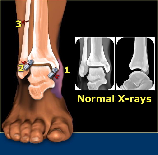
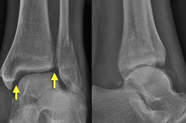
Non Displaced Medial Malleolar Fracture Image Radiopaedia Org In that case we have the following combination: stage 1: rupture of the medial collateral ligament stage 2: rupture of the anterior syndesmosis. stage 3: high fibular fracture. stage 4: tertius fracture. an isolated tertius fracture on the ankle radiographs indicates the presence of an unstable ankle injury. Weber b fractures could be further subclassified as 9. b1: isolated. b2: associated with a medial lesion (malleolus or ligament) b3: associated with a medial lesion and fracture of posterolateral tibia. type c. above the level of the syndesmosis (suprasyndesmotic) tibiofibular syndesmosis disruption with widening of the distal tibiofibular. Citation, doi, disclosures and article data. ankle fractures account for ~10% of fractures encountered in trauma, preceded only in incidence by proximal femoral fractures in the lower limb. they have a bimodal presentation, involving young males and older females. ankle injuries play a major part in functional impairment after multi or. An isolated fracture of the medial malleolus, or widening of the ankle joint with no visible fracture seen on ankle x ray, should raise the suspicion of an associated fracture of the fibula. if this is not visible in the distal fibula then further x rays of the proximal fibula should be performed. imaging of the proximal fibula should also be.

The Radiology Assistant Ankle Fracture Mechanism And Radiography Citation, doi, disclosures and article data. ankle fractures account for ~10% of fractures encountered in trauma, preceded only in incidence by proximal femoral fractures in the lower limb. they have a bimodal presentation, involving young males and older females. ankle injuries play a major part in functional impairment after multi or. An isolated fracture of the medial malleolus, or widening of the ankle joint with no visible fracture seen on ankle x ray, should raise the suspicion of an associated fracture of the fibula. if this is not visible in the distal fibula then further x rays of the proximal fibula should be performed. imaging of the proximal fibula should also be. Lateral malleolar fracture. isolated lateral malleolar fractures are common. the weber classification is used to determine treatment. weber a: below the ankle joint with intact syndesmosis. weber b: at the level of the ankle joint weber c: above the ankle joint with medial malleolus fracture. more: lateral malleolar fracture. maisonneuve fracture. The ankle is a hinge joint formed by three bones: the tibia, the fibula and the talus. proximally, the joint comprises the medial malleolus (the distal end of the tibia), the tibial plafond and the lateral malleolus (the distal end of the fibula) which collectively form a rectangular socket called the mortise into which fits the talar dome.

The Radiology Assistant Algoritm For Ankle Fractures 2 0 Lateral malleolar fracture. isolated lateral malleolar fractures are common. the weber classification is used to determine treatment. weber a: below the ankle joint with intact syndesmosis. weber b: at the level of the ankle joint weber c: above the ankle joint with medial malleolus fracture. more: lateral malleolar fracture. maisonneuve fracture. The ankle is a hinge joint formed by three bones: the tibia, the fibula and the talus. proximally, the joint comprises the medial malleolus (the distal end of the tibia), the tibial plafond and the lateral malleolus (the distal end of the fibula) which collectively form a rectangular socket called the mortise into which fits the talar dome.

Comments are closed.